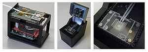
The ChipScope project has developed the scientific and technological basis for a completely new microscopy approach to optical super-resolution. The project, coordinated by the University of Barcelona, has realised semiconductor nano-light-emitting diode (nanoLED) arrays for this purpose. In the future, this is expected to revolutionise optical microscopy, making it chip-sized, convenient, affordable and ubiquitously available - not only for laboratories but also for everyday life. Experts of the Competence Unit Molecular Diagnostics, Center for Health & Bioresources, have developed a sample handling system that transports the samples to the desired target position and supplies the cells seeded in the microfluidic channels with nutrients. ChipScope ran for four years until the end of 2020 and was funded by the European Union's H2020 research and innovation programme with almost €4 million (grant agreement number 737089).
The EU-funded ChipScope project, with the participation of the Competence Unit Molecular Diagnostics, Center for Health & Bioresources, was successfully completed in December 2020. An interdisciplinary team of researchers from five European institutions explored a completely new strategy for optical microscopy.
Stefan Schrittwieser, project coordinator at AIT explains "In classical optical microscopy, the examined sample area is illuminated simultaneously and the light scattered from each point is collected with an area-selective detector, e.g. the human eye or the sensor of a camera." This is different with ChipScope, Schrittwieser explains further "With the ChipScope idea, a structured light source with tiny, individually addressable elements is used instead. The specimen is located in close proximity above this light source. Whenever individual emitters are activated, the light propagation depends on the spatial structure of the specimen, much like what is known in the macroscopic world as a shadow image. To obtain an image, the total amount of light transmitted through the sample region is detected by a detector that activates one light element at a time, scanning the sample space." The light elements have sizes in the nanometre range, the image is thus captured part by part and reassembled by software to form the final image - by breaking this down to the smallest areas, a super-resolution result is possible.
To realise this completely new idea, a bundle of newly developed technologies was necessary. The structured light source was realised by nanometre-sized light-emitting diodes (LEDs), which were developed at the Technical University of Braunschweig. LEDs with a diameter of 200 nm were produced as an array with a distance of 200 nm between the LEDs. For the microscope prototype, the University of Barcelona, which also coordinated the project, developed electronic components to directly control each LED, highly sensitive light detectors and a custom imaging software.
One of the main challenges was to bring the samples, human lung cells, close to the structured light source. This is where the AIT experts contributed their expertise and developed a microfluidic sample handling system for this purpose, in which a fine system of channels was integrated into a polymer matrix. This enables the sample to be transported through a fluid to the target position and supplies the cells seeded in the microfluidic channels with nutrients.
Other partners in the ChipScope project include a team from the Medical University of Vienna, the University of Rome Tor Vergata, Ludwig Maximilians University in Munich and FSRM, Switzerland. This project was funded by the European Union's H2020 research and innovation programme (grant agreement number 737089).
Project homepage: http://www.chipscope.eu/
Project video: https://www.youtube.com/watch?v=BxfKJeHU33I
More information: https://cordis.europa.eu/project/id/737089


